
1 / 5


| Model NO. | V260 | Condition | New |
| Using Ambient | Hospital | Certifications | ISO13485, ISO14001, ISO45001, ISO9001 |
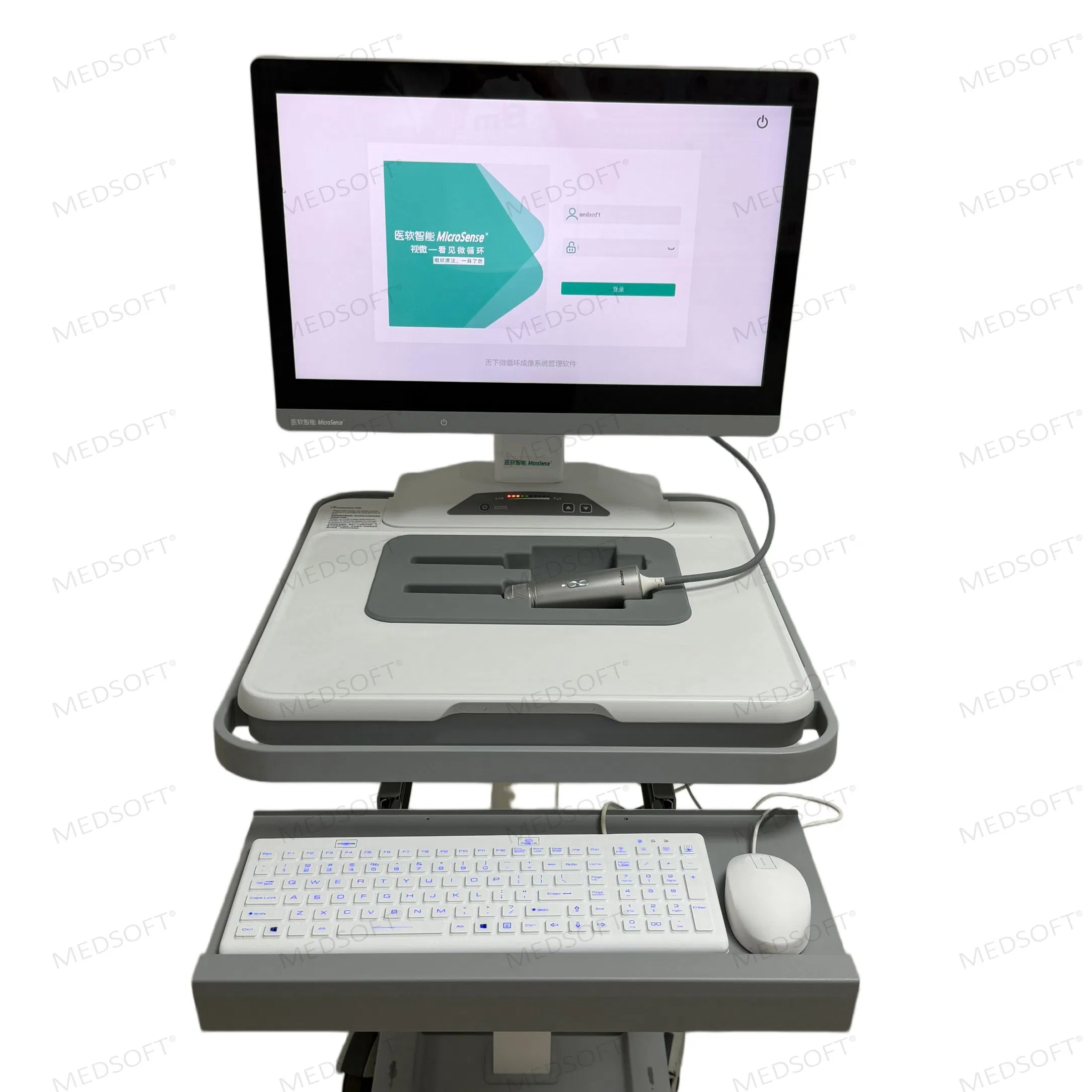
| Name | Sublingual Microcirculation Imaging System | Samples | Available |
| OEM/ODM | Available | Service | Lifetime Support |
| User Manual | Available | Factory Experience | Over 17 Years |
| Length | 135.2mm±5mm | Transport Package | Carton/Wooden Box |
| Production Capacity | 1200 PCS/Year | HS Code | 90111000 |

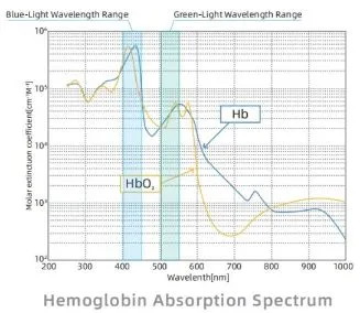
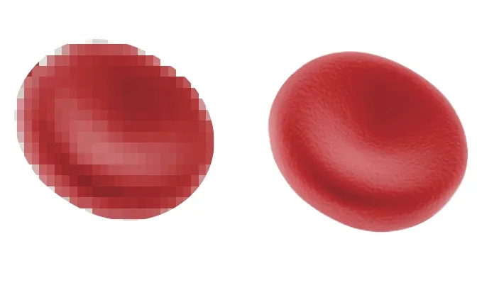
According to the principles of optical physics, resolution (r) is defined as λ / (2NA). With a fixed numerical aperture (NA), a shorter wavelength (λ) results in a smaller resolution value (r), thereby enabling superior visualization of minute structures such as individual red blood cells.
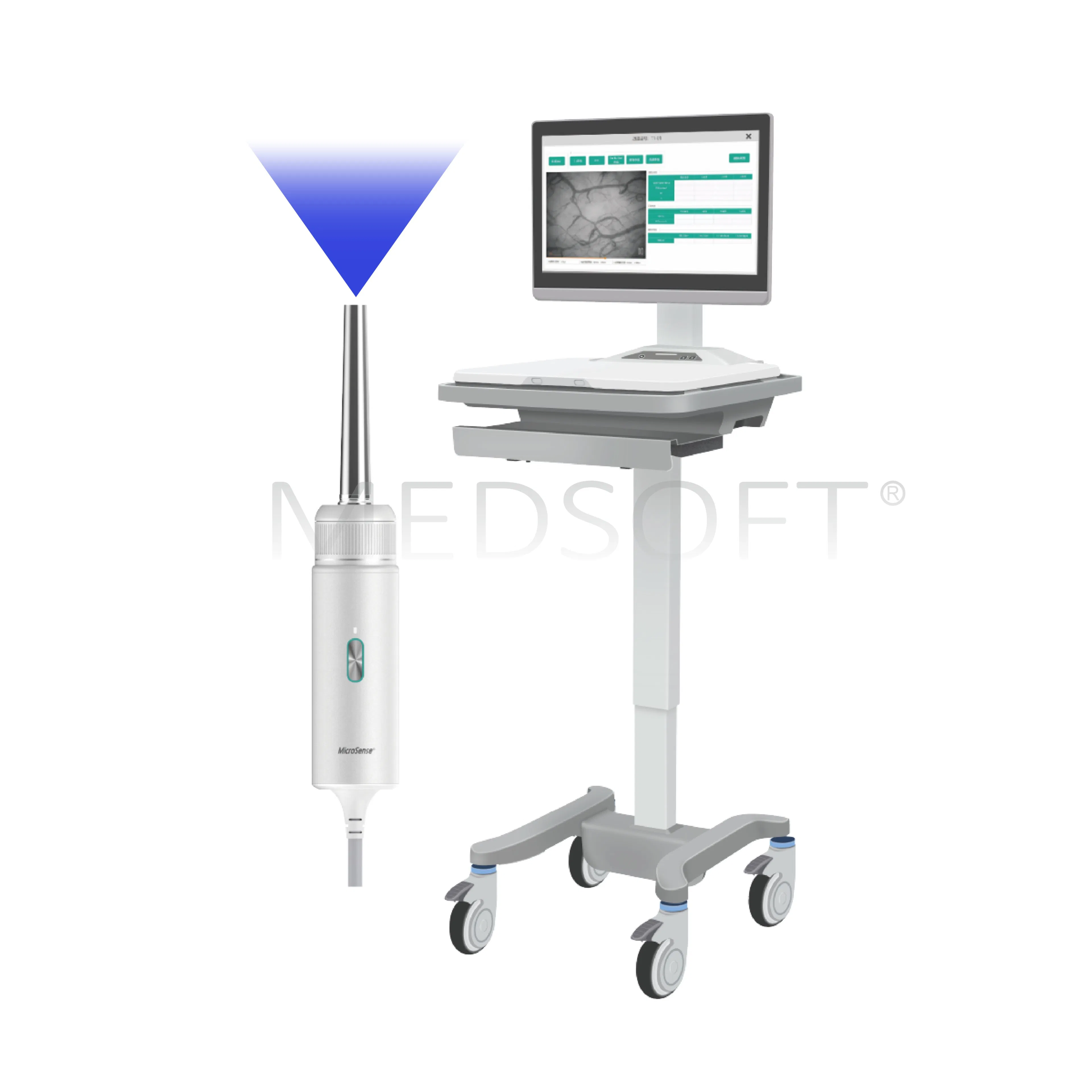
The V260 is the first sublingual microcirculation imaging system to adopt blue-light imaging. Compared with conventional green-light imaging, it achieves nanometer-level resolution, enabling sharper and more precise visualization of individual red blood cells.
Hemoglobin absorption peaks at 415 nm (blue-light). Thus, V260 achieves high-contrast, crystal-clear images, enabling precise bedside assessment of sublingual microcirculation and tissue perfusion.
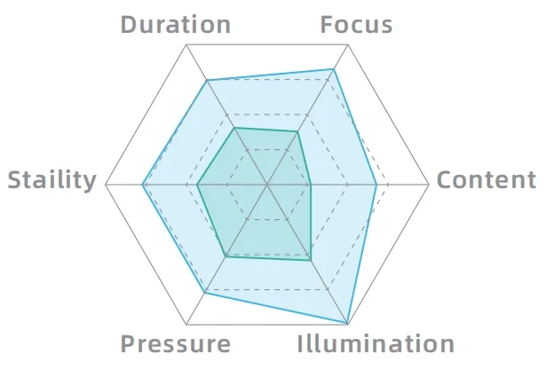
With equal observation areas, blue-light (415 nm) reveals significantly more microvessels compared to green-light (520 nm), allowing for improved evaluation of RBC flow (TVD: 24.8±3.1 vs. 20.0±3.0, P<0.0001).
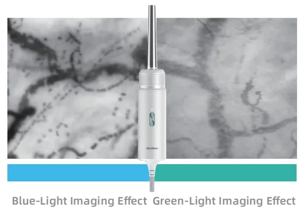
Based on the Microcirculatory image quality scoring (MIQS), the acceptability of blue-light (415 nm) imaging quality is 1.5 times higher than that of green-light (520 nm) (P < 0.001).
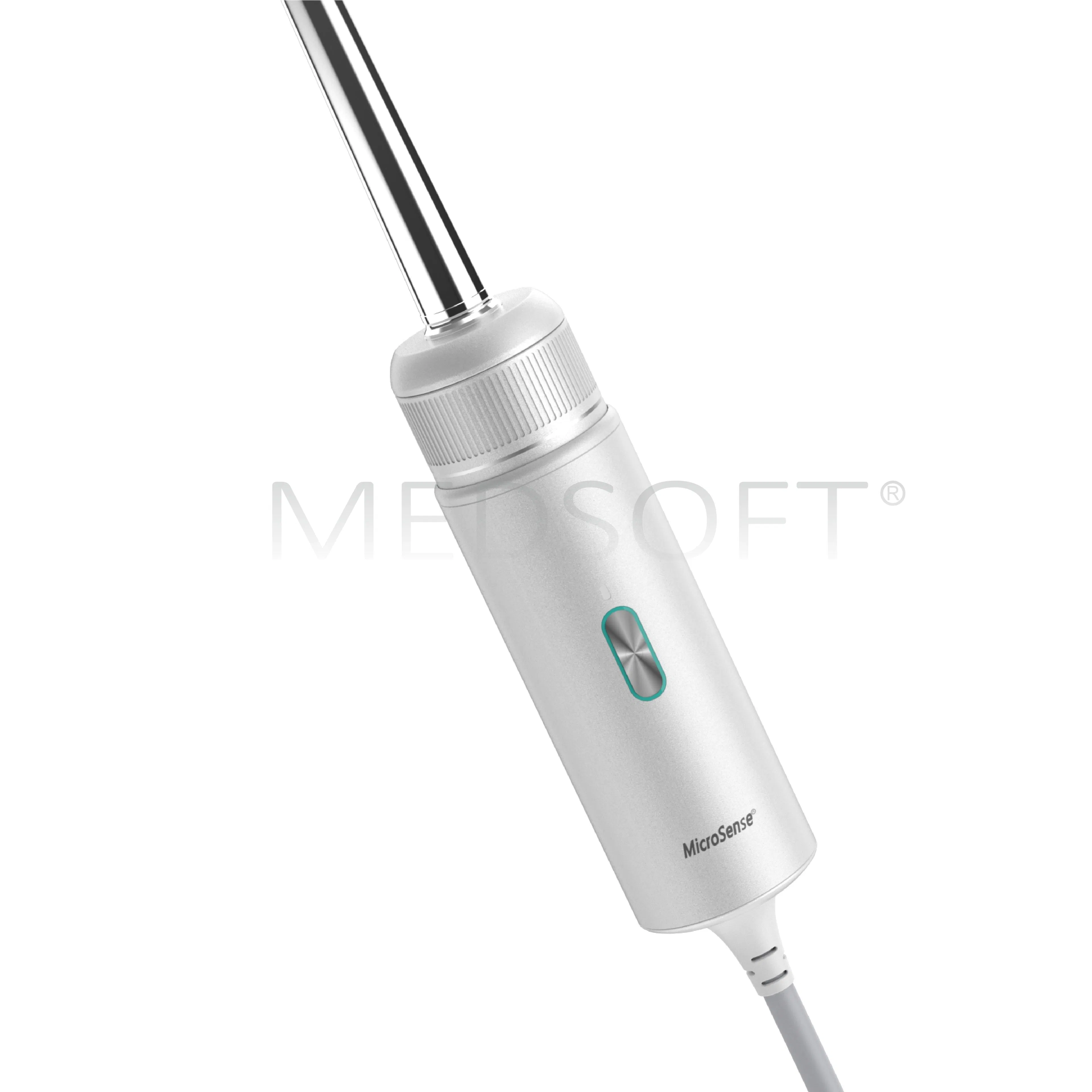
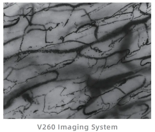
Visualizes red blood cells and intercellular spaces with exceptional clarity. While maintaining a slim probe diameter and incorporating a high-resolution camera, the V260 achieves an optical resolution below 1 um.
Reduces heterogeneity in the observed field, enabling more comprehensive analysis of microcirculatory regions. Combining ultra-high optical resolution with 4X magnification yields an expanded effective observation area.

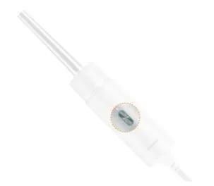
Eliminates red blood cell (RBC) motion artifacts, even under high-dynamic conditions. The field of view is illuminated by ten LED light sources with incident-angle strobe lighting. With a strobe duration < 1ms and 60 fps recording, motion artifacts are effectively eliminated.

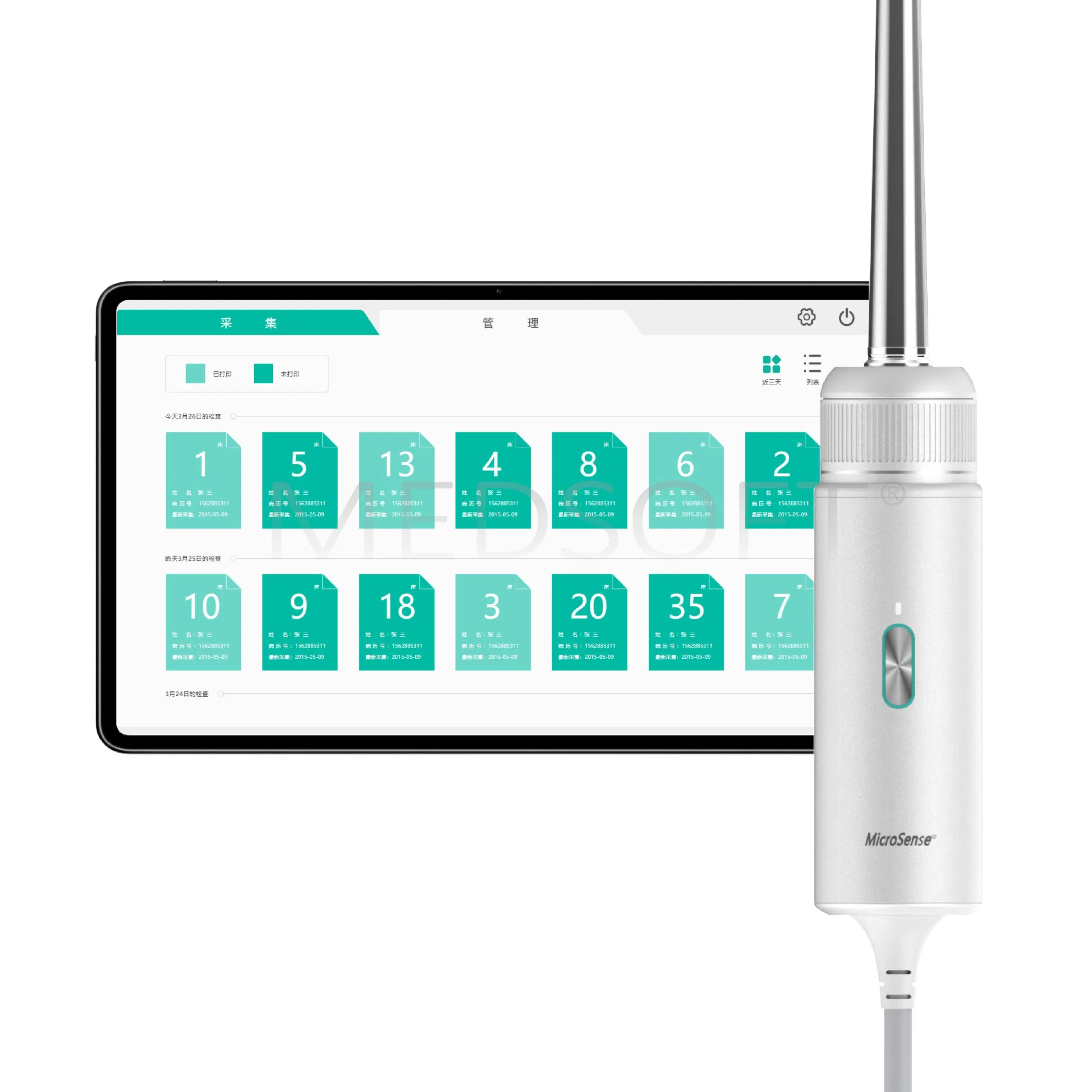
Effortless image acquisition by a single operator. The handle integrates a one-button start/stop recording function, enabling smooth single-hand operation without additional keyboard or touchscreen input.
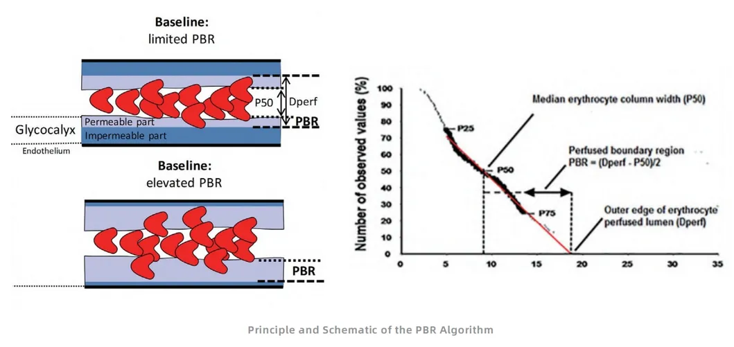
The endothelial glycocalyx functions as a protective barrier of vascular integrity. Damage results in increased vascular permeability, aggravated inflammation, and altered vascular tone. Syndecan-1 is a pivotal biomarker of glycocalyx degradation.
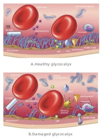
Patients with sepsis or septic shock may experience glycocalyx degradation, manifested as an increased Perfusion Boundary Region (PBR). The V260 integrates the PBR algorithm for rapid bedside assessment.
| De Backer score | Total Vascular Density (TVD) |
| Small Vessel Density (sTVD) | Proportion of Perfused Vessels (PPV) |
| Proportion of Perfused Small Vessels (sPPV) | Perfused Vessels Density (PVD) |
| Perfused Small Vessels Density (sPVD) | Microvascular Flow Index (MFI) |
| Heterogeneity Index (HI) | Red Blood Cell velocity (RBCv) |
| Perfused Boundary Region (PBR) | - |


Professional Expertise: Focus on R&D of Medical Software and Medical Devices in Critical Care Medicine. Dedicated to transforming clinical needs into innovative products that enhance patient survival rates.
Product Portfolio: