1 / 5
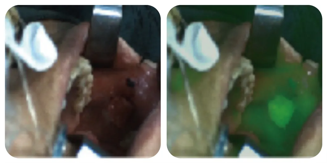
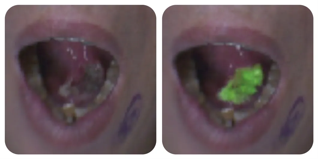
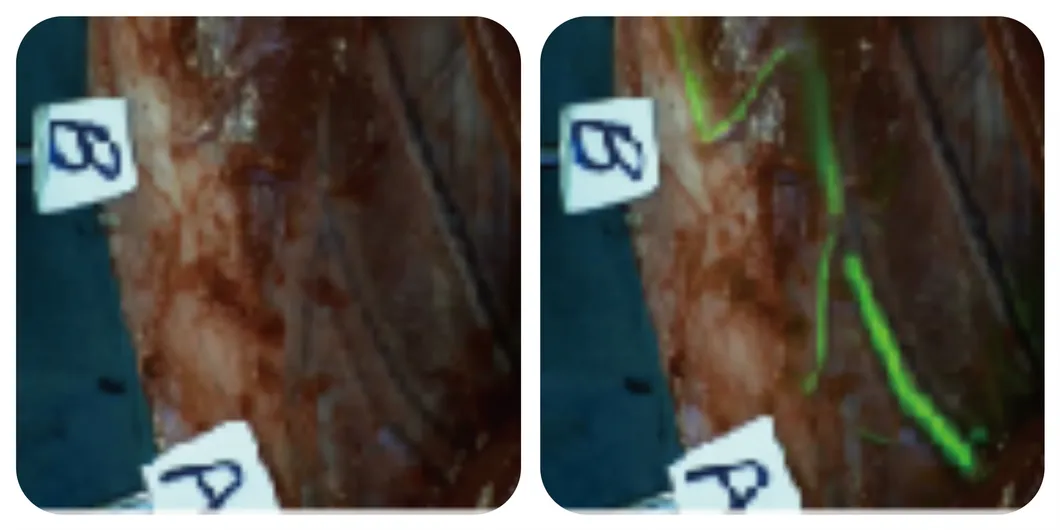
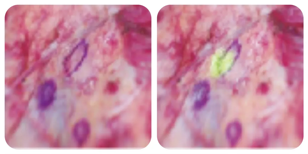
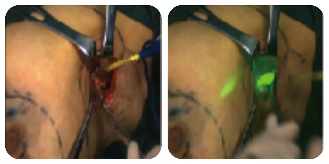
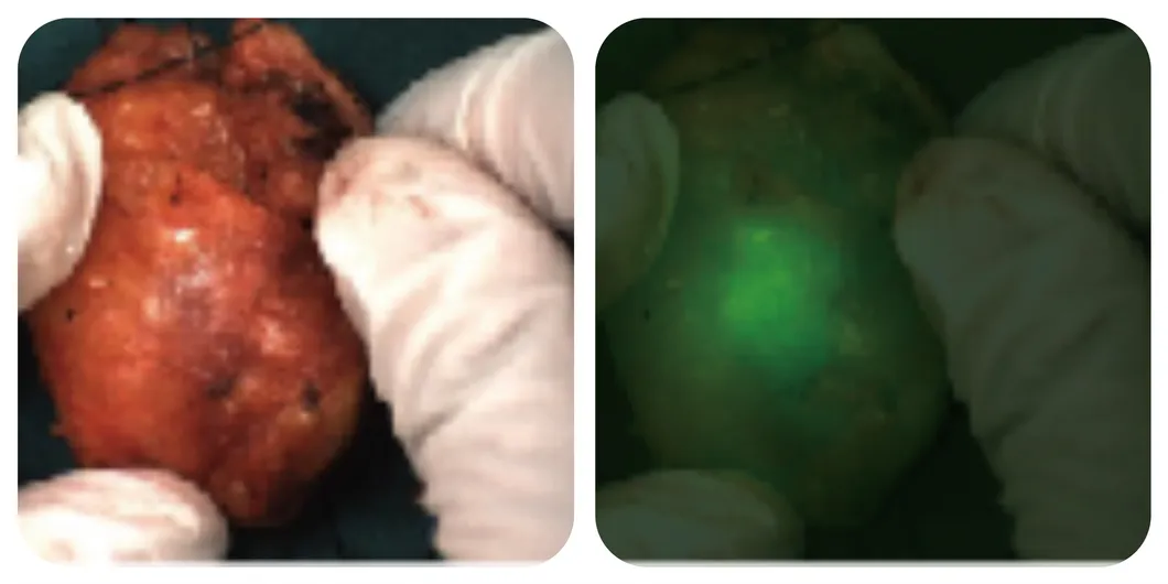
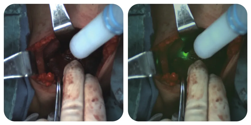
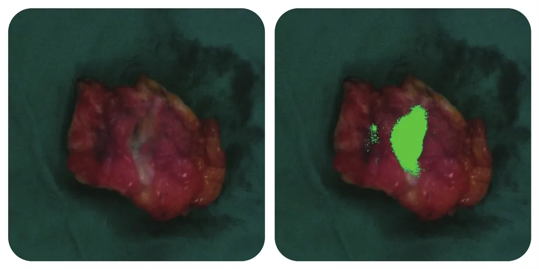
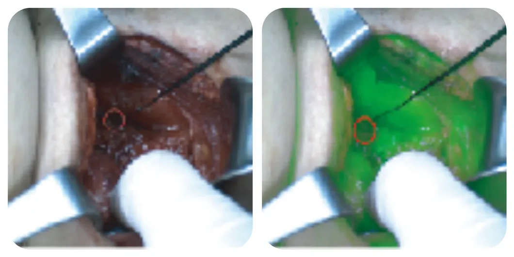
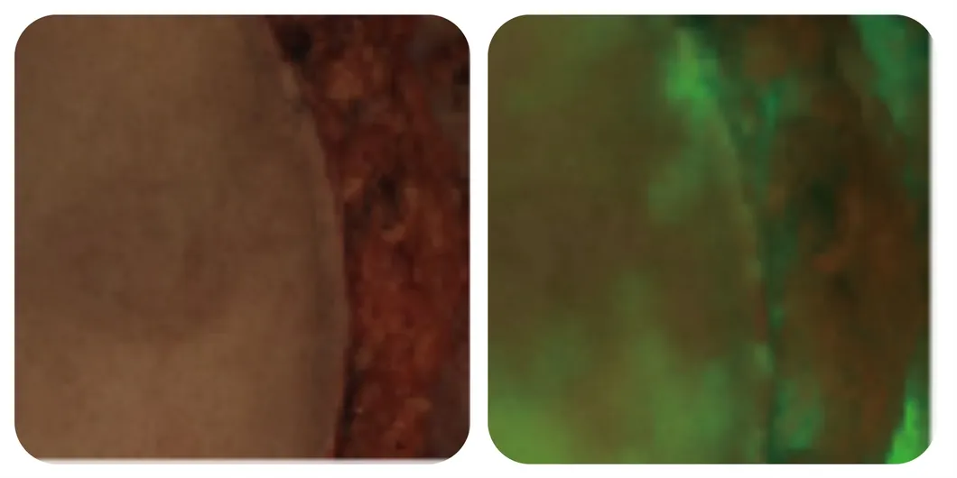
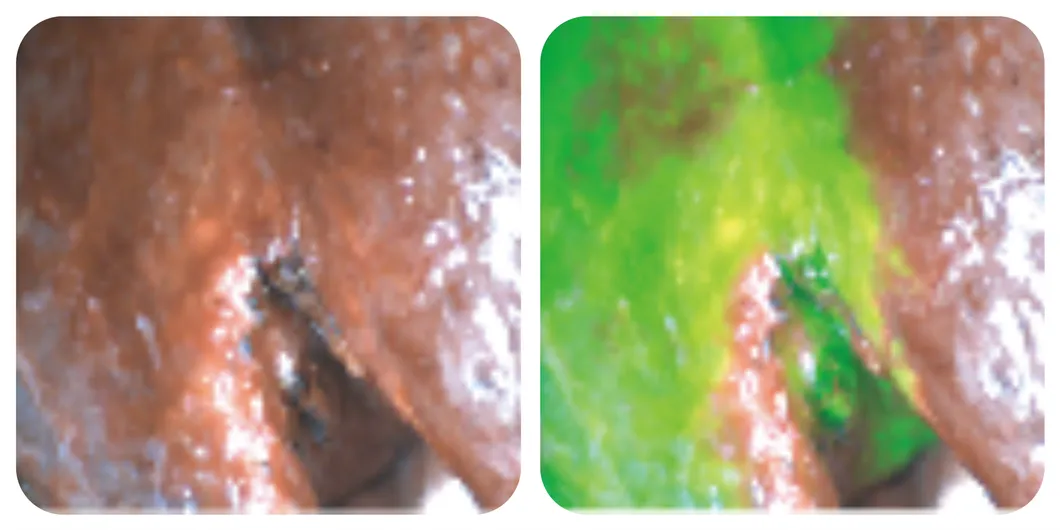
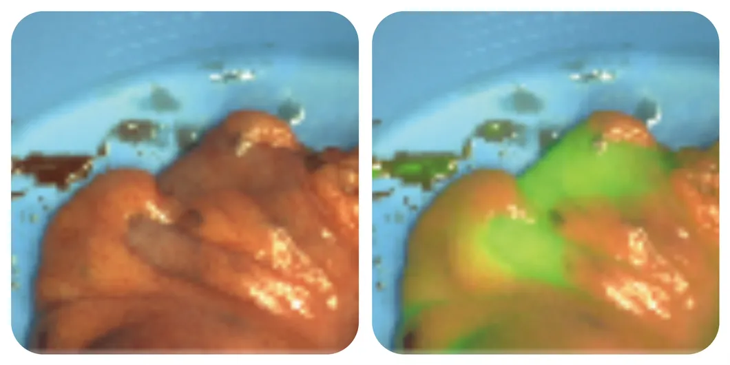
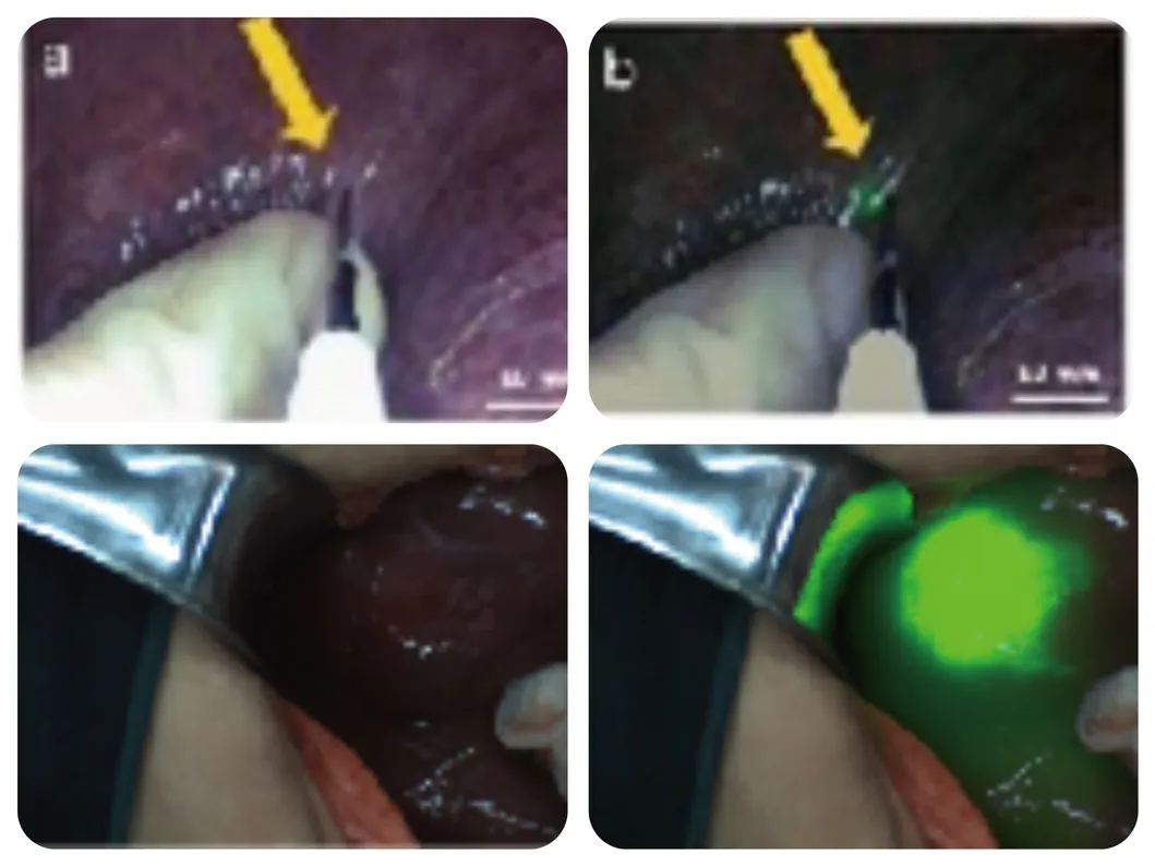
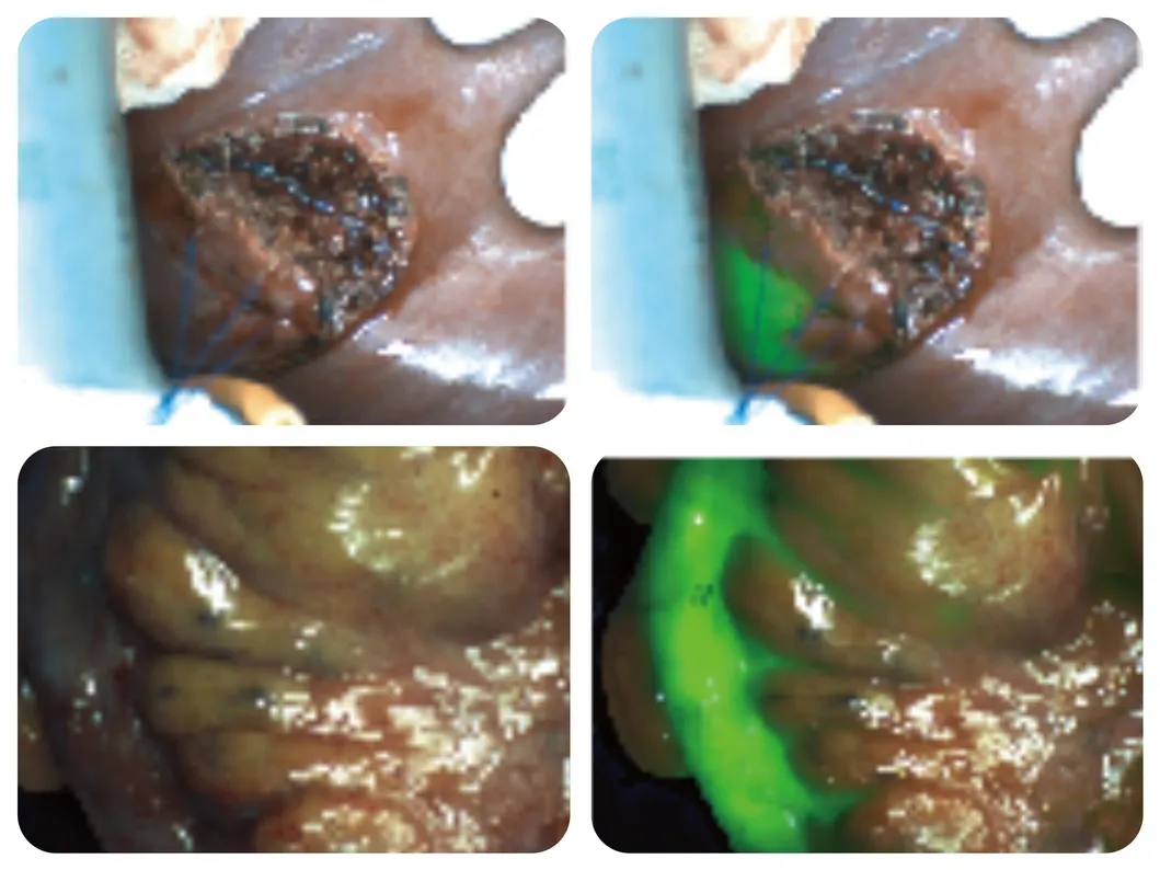
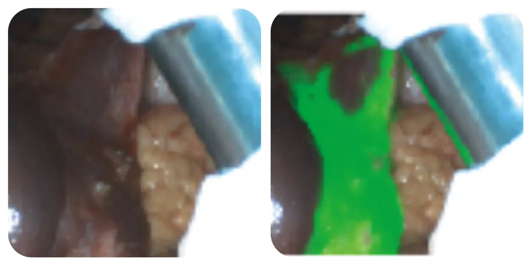
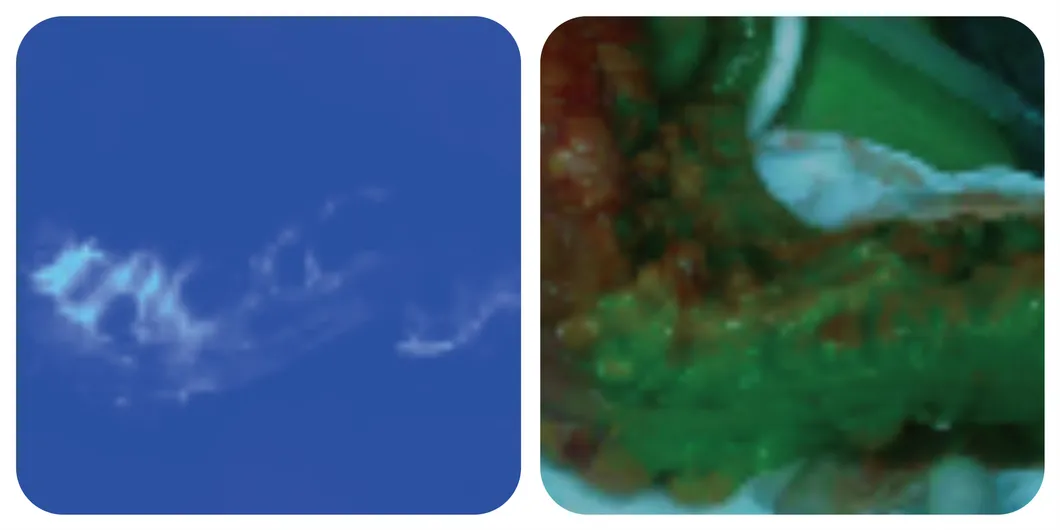
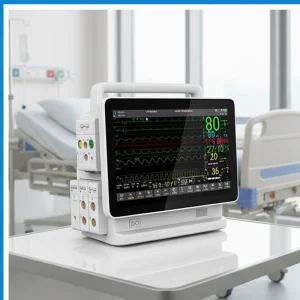
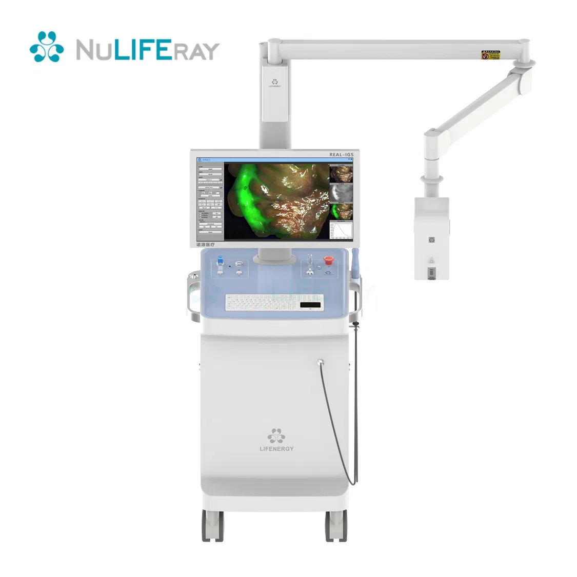
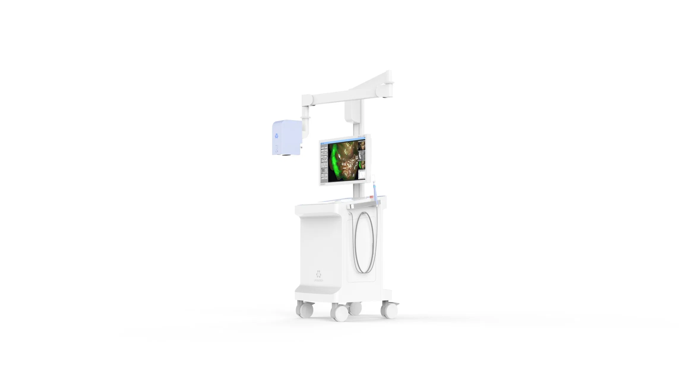
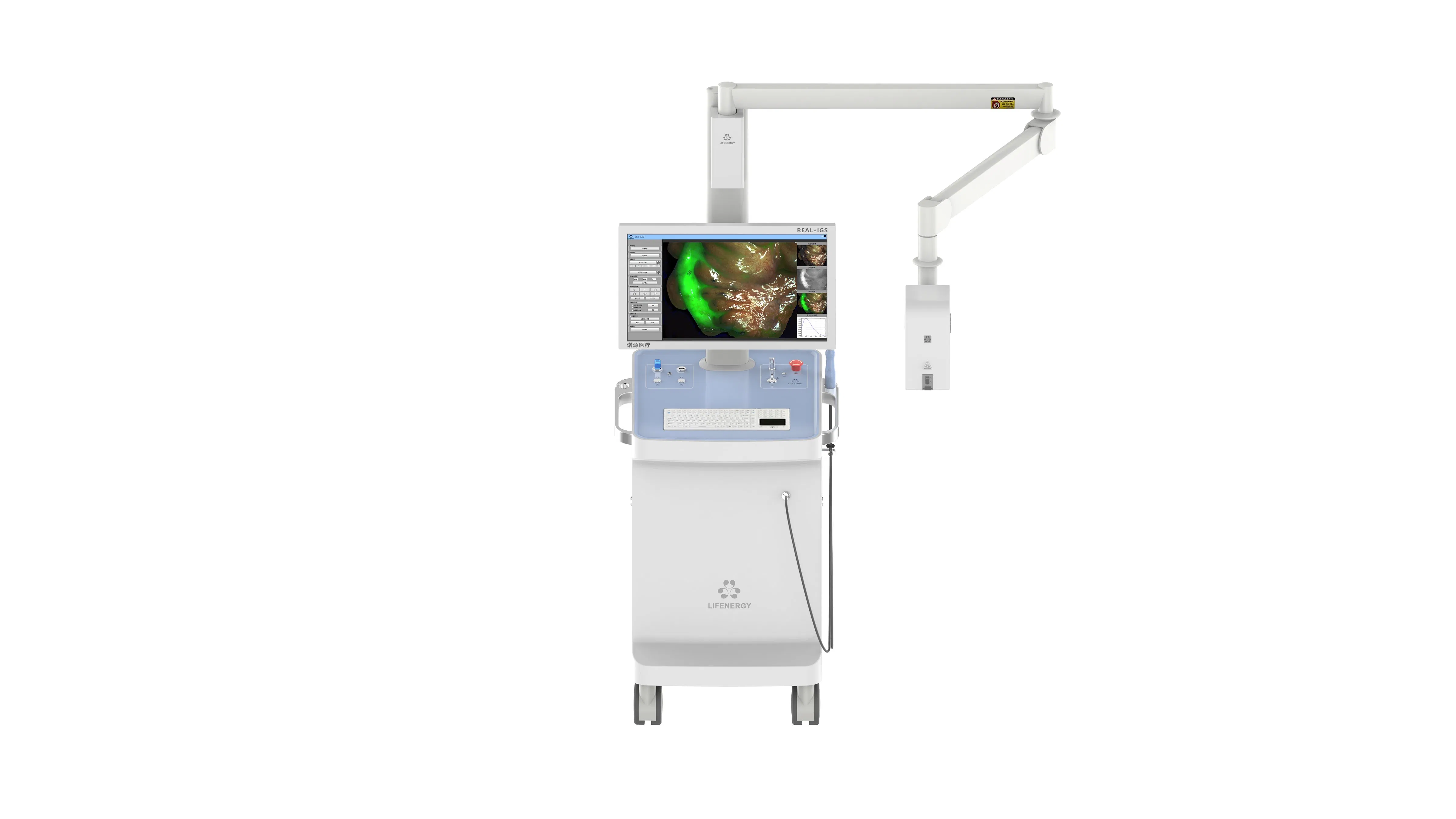
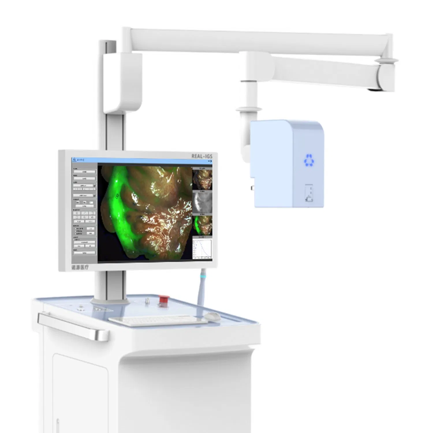
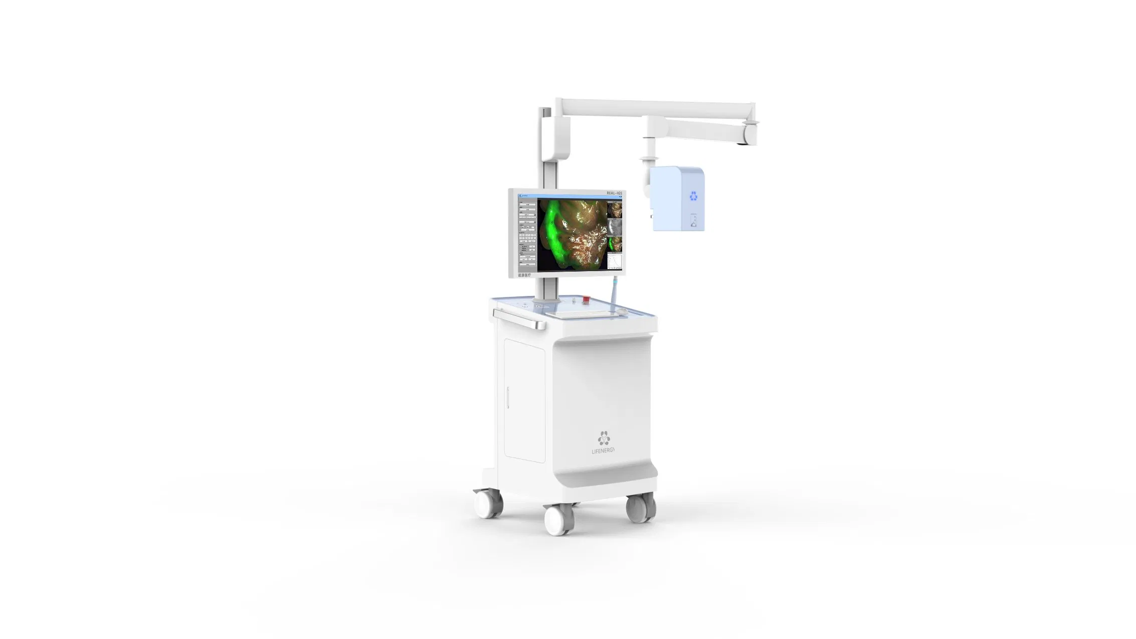
FL1-10B Surgical Fluorescence Imaging System is a surgical guidance system that uses a drug-device combination approach. It employs indocyanine green (ICG) as a fluorescence probe, relying on "ultra-high sensitivity" and combining the optical properties of ICG in submillimeter-size tumors to provide surgeons with high-definition visible light, fluorescence imaging, and quantitative data for diagnostic information during tumor surgery.
It is suitable for real-time observation of tissues (such as tumor tissue, margin tissue), blood supply (free skin flap), lymph nodes (sentinel lymph nodes, regional lymph nodes), and anatomical structures (liver segments, gallbladder, lung segments). This allows for more accurate medical judgments, optimized surgical plans, and evaluated treatment efficacy across multiple clinical departments, including oral and maxillofacial surgery, plastic and reconstructive surgery, thyroid surgery, breast surgery, pediatric surgery, and general surgery.
| Project | Content |
|---|---|
| Number of Camera Chips | 2CMOS |
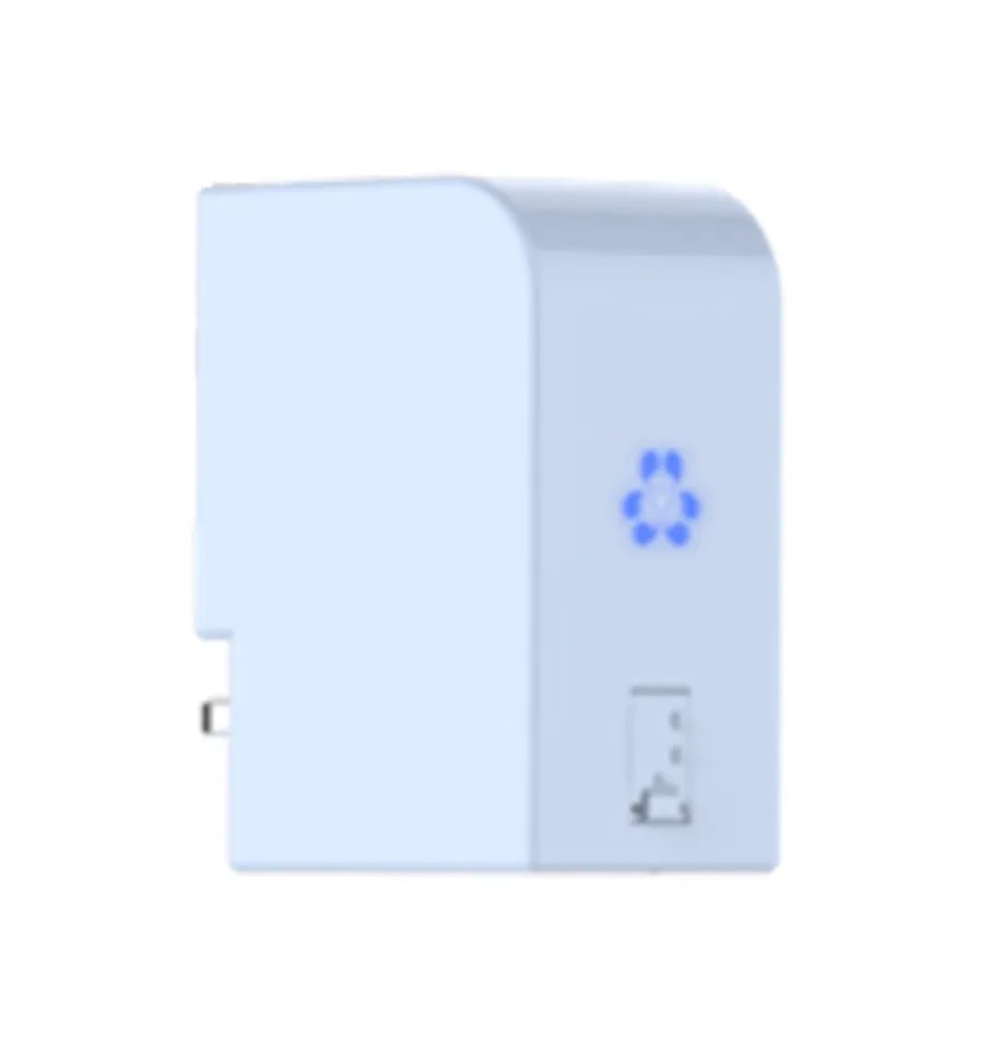
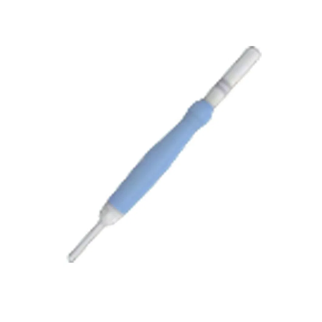
| Convenient Handheld Probe | Handheld Spectral Quantitative Analysis Probe |
| Special Fluorescence Development | AI Assisted Boundary Sharpening |
| Image Mode | 7 Types: White Light/Fluorescence/Fusion/Multimode/ColorGrading/Quantification/Spectroscopy |
| Laser Wavelength | 785nm |
| Workstation Available | Yes |
| Workstation Function | Fluorescence Intensity & Spectral Quantitative Analysis |
| Fluorescence Detection Limit | 10-12M/L |
| Lens Zoom Factor | 4 Times |
| Camera Working Distance | Recommended 10-25cm |
| Focus Mode | Electric Focus |
| White Balance Method | Manual White Balance |
| Laser Grade | 3R |
| Integrated Model | Yes |
| Recording System Resolution | High Definition |
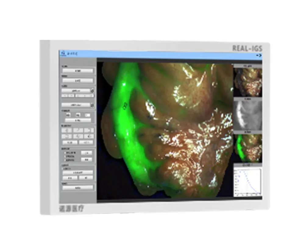
Used in different surgical procedures with the fluorescent contrast agent indocyanine green (ICG), it displays the circulation of the lymphatic system and blood vessels, as well as the perfusion of related tissues through near-infrared fluorescence imaging technology.